Sketch And Label Of A Cross Section Of A Long Bone - Labeling portions of a long bone learn with flashcards, games and more — for free.. A = epiphysis b = diaphysis c = articular cartilage d = periosteum f = compact bone g = medullary cavity (yellow marrow) h = endosteum j = epiphyseal line (growth plate). A hard outer layer that is dense, strong, and durable. The compact bone is made up of osteon. It suggests that the bone will have equal strength in all directions. The 2020 pandemic required that many of these activities be converted a format that students could complete online with a device, making labeling and coloring a little tricky.
Anatomycorner is a branch of biologycorner.com focused on dissections and body systems. Diaphysis elongated hollow central portion of the bone located between the methaphyses. Epiphysis • the two ends of a long bone which are wider than the shaft and take part in the formation of a joint b. You can specify conditions of storing and accessing cookies in your browser. Observed 2.sketch and label the diaphysis of the beef bone:
Plate 3.49: Bone from www.anatomyatlases.org A = epiphysis b = diaphysis c = articular cartilage d = periosteum f = compact bone g = medullary cavity (yellow marrow) h = endosteum j = epiphyseal line (growth plate). The outer layer is usually hard and compact, and it covers the inner spongy cavity containing the bone marrow. You can specify conditions of storing and accessing cookies in your browser. Diaphysis elongated hollow central portion of the bone located between the methaphyses. The head of each end of a long bone consists largely of spongy bone and is covered with hyaline cartilage. Long bones are covered by a membranous layer called the periosteum. Epiphyseal disc • in the embryo and the growing child it is a cartilaginous plate located between the epiphysis and the. All photography, text, and labels by rob swatski, assistant professor of biology, harrisburg.
Labeling portions of a long bone learn with flashcards, games and more — for free.
Two types of bone tissues in cross section of a long bone : The head of each end of a long bone consists largely of spongy bone and is covered with hyaline cartilage. Bones are also very good at repairing themselves. This quiz on human bones is designed to test your knowledge on the location of each individual bone. The length of the bone forms the main shaft. This is page 4 of a photographic atlas i created as a laboratory study resource for my biol 121 anatomy and physiology i students on the bones and bony landmarks of the appendicular skeleton. A special exihibition in the centre historique minier in lewarde featuring a large number of models on mining in the 20th century. From wikimedia commons, the free media repository. Terms in this set (12). Solved correctly label the following anatomical parts of. It's not impossible, but if students get frustrated trying to use apps to complete assignments, teachers find that many will give up. The outer layer is usually hard and compact, and it covers the inner spongy cavity containing the bone marrow. Diaphysis elongated hollow central portion of the bone located between the methaphyses.
Labeling portions of a long bone learn with flashcards, games and more — for free. Refer to as you study the following section. Model of a coal mine. Epiphyseal disc • in the embryo and the growing child it is a cartilaginous plate located between the epiphysis and the. All photography, text, and labels by rob swatski, assistant professor of biology, harrisburg.
Pin by Danielle Papas on Nursing Anatomy & Physiology ... from i.pinimg.com They consist of 2 heads proximal and distal epiphysis respectively, connected with the a long bone like a femur in the thigh is commonly used as an example of a bone in the human body. You can specify conditions of storing and accessing cookies in your browser. Sketch a typical long bone and label its epiphyses diaphysis. The diagram depicts a cross section of three different kinds of mines, along with structural elements of the underground shaft and slope mines. Anatomycorner is a branch of biologycorner.com focused on dissections and body systems. Soft, porous and can retain more water. A uniform cross section is the cross section of the solid, parallel to base, such that the resulting figure has the same shape and size as that of the base of the figure.more about uniform cross sectionsolids like pyramids and. All photography, text, and labels by rob swatski, assistant professor of biology, harrisburg.
It's not impossible, but if students get frustrated trying to use apps to complete assignments, teachers find that many will give up.
The end of a growing tibia, cut lengthwise*. This quiz on human bones is designed to test your knowledge on the location of each individual bone. Anatomycorner is a branch of biologycorner.com focused on dissections and body systems. It's not impossible, but if students get frustrated trying to use apps to complete assignments, teachers find that many will give up. A uniform cross section is the cross section of the solid, parallel to base, such that the resulting figure has the same shape and size as that of the base of the figure.more about uniform cross sectionsolids like pyramids and. Examples and applications of a datum planes and how and when and why you should use them in freecad. Describe the tissues you observedquestions:a.how does the model of the femur compare to the diagrams in your textbook or this manual?b.how does the texture of articular cartilage compare to that of periosteum?c.what is. In your anatomy & physiology lecture and lab class, you will be required to name each individual bone in the human body. Terms in this set (12). With examples of use slanted features, sketch on curve, cross section. You can specify conditions of storing and accessing cookies in your browser. Labeling portions of a long bone. This site is using cookies under cookie policy.
Describe the tissues you observedquestions:a.how does the model of the femur compare to the diagrams in your textbook or this manual?b.how does the texture of articular cartilage compare to that of periosteum?c.what is. This quiz on human bones is designed to test your knowledge on the location of each individual bone. A hard outer layer that is dense, strong, and durable. A uniform cross section is the cross section of the solid, parallel to base, such that the resulting figure has the same shape and size as that of the base of the figure.more about uniform cross sectionsolids like pyramids and. You can specify conditions of storing and accessing cookies in your browser.
Figure 10. General structure of a mammalian bone. Other ... from openi.nlm.nih.gov Size of this png preview of this svg file: Diagram of transverse section of a mammalian bone. Diaphysis elongated hollow central portion of the bone located between the methaphyses. The head of each end of a long bone consists largely of spongy bone and is covered with hyaline cartilage. All photography, text, and labels by rob swatski, assistant professor of biology, harrisburg. Describe the tissues you observedquestions:a.how does the model of the femur compare to the diagrams in your textbook or this manual?b.how does the texture of articular cartilage compare to that of periosteum?c.what is. It has a long central area called a shaft or diaphysis and. A hard outer layer that is dense, strong, and durable.
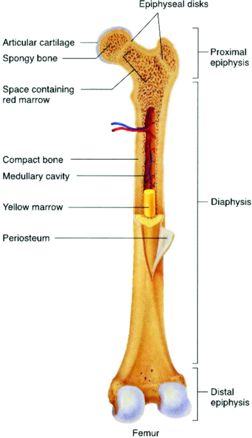
The femur, the bone of the thigh, will be used as an example in considering the structure of a long bone.
The head of each end of a long bone consists largely of spongy bone and is covered with hyaline cartilage. This site is using cookies under cookie policy. Diagram of transverse section of a mammalian bone. A = epiphysis b = diaphysis c = articular cartilage d = periosteum f = compact bone g = medullary cavity (yellow marrow) h = endosteum j = epiphyseal line (growth plate). As a nurse, you will need to know the basic about the human skeleton. In this lab you can explore the bones of the human skeleton using our skeleton viewer that can also be played as a game. Hard and difficult to dig. Soft, porous and can retain more water. The compact bone is made up of osteon. It's not impossible, but if students get frustrated trying to use apps to complete assignments, teachers find that many will give up. The end of a growing tibia, cut lengthwise*. A hard outer layer that is dense, strong, and durable. Diaphysis elongated hollow central portion of the bone located between the methaphyses.


0 Komentar